

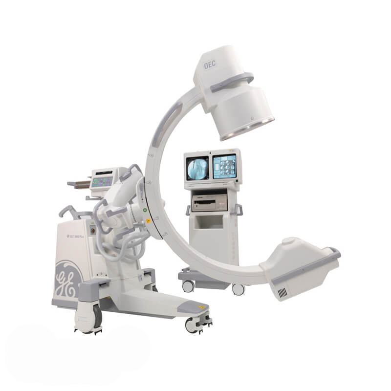

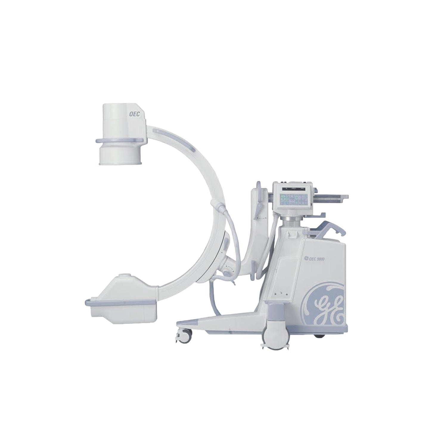
The GE OEC 9600 is a mobile x-ray system used for various diagnostic and surgical applications. It provides high quality fluoroscopic imaging and film radiographs.
PDF Brochure
Features & Specifications
• Dual Hi-Resolution 16” Square Monitors
• Rotating X-ray Tube
• Tri-Mode 9″/6″/4 Image Intensifier (II)
• High Resolution CCD
• 360° motorized rotation - video camera
• On-screen orientation indicator (real-time feedback without fluoro)
• Negative mode for camera
• Left-right image reversal
• Top-bottom image reversal
• Fluoro Boost Mode
• Pulsed Fluoro Mode
• General Surgical Package (GSP)
• Image Storage
• Last Image Hold (LIH)
• MARS-Motion Artifact Reduction System
• Gamma correction
• Digital windowing
• Patient annotation
• DICOM
• Entire system is computer controlled. Software upgrade-able
• Hand-held X-Ray remote control
• Multi-function footswitch
• Multi-function infrared remote control
• Monitor Cart Dimensions: 27-1/3″ D x 27-1/3″ W x 64-1/3″ H
• C-Arm Frame Dimensions: 76″ L x 32-1/3″ W x 67-2/3″ H
• Source to image distance: 39 inches
• Free space in arc: 31 inches
• Depth in arc: 26 inches
• Arc orbital movement: 9 inch image intensifier – 115 degrees
• Arc orbital movement: 12 inch image intensifier – 112 degrees
• Left / Right wig-wag scan: ± 11º
• Vertical travel: 18 inches motorized
• Horizontal travel: 8 inches
• L-Arm rotation: ±180 degrees motorized
• Reversible C-Arm: 180 degrees manual (flip-flop)
• Video signal: Standard RS 170A 60 Hz, 525 lines
• Video Camera Aspect ratio: 4:3
Modes
Fluoroscopy Mode
• Focal spot: 0.3 mm
• kVp range: 40 – 120 kVp
• mA range: 0.2 – 5.0 mA normal mode
• mA range: 1.0 – 12 mA continuous fluoro boost mode
• Auto and manual modes
• Continuous, one-shot or pulsed operation
• ABS varies mA, kVp and camera gain
• User specific ABS tables
Pulsed Fluoroscopy Mode
• Focal spot: 0.3 mm
• kVp range: 40 – 120 kVp
• mA range: 0.2 – 5.0 mA
• Pulse rate: 1, 2, 4, or 8 pulses per second
• Pulse width: 30 or 50 milliseconds
• Computer controlled ABS, mA, kVp and camera gain
Radiographic Mode
• Focal spot: 0.3 mm or 0.6 mm
• Focal spot automatically selected
• 0.3 mm – mAs range: 1 – 100 mAs
• 0.6 mm – mAs range: 110 – 300 mAs
• kVp range: 50 – 120 kVp